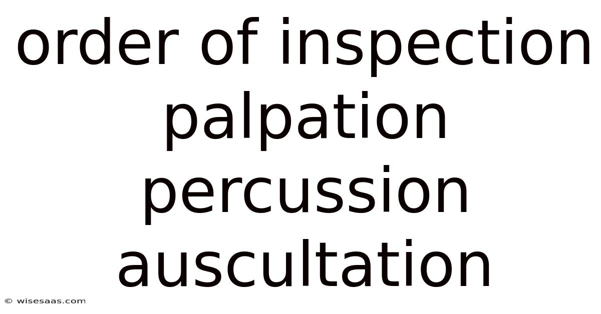
The Order of Inspection, Palpation, Percussion, and Auscultation: A Systematic Approach to Physical Examination
In clinical medicine, the physical examination is a foundational skill that allows healthcare providers to gather critical information about a patient’s health status. Now, this structured approach ensures that each step builds on the previous one, minimizing the risk of missing subtle findings and maximizing diagnostic accuracy. Which means among the most essential techniques in this process are inspection, palpation, percussion, and auscultation—four methods that, when performed in a specific sequence, enable clinicians to assess organ function, detect abnormalities, and guide diagnostic decisions. Understanding the rationale behind this order is vital for medical students, nurses, and practitioners alike, as it forms the backbone of effective patient evaluation.
Step 1: Inspection – The Visual Assessment
The physical examination begins with inspection, a non-invasive technique that relies on visual observation. This step allows clinicians to gather preliminary information about a patient’s general appearance, skin integrity, posture, and any visible abnormalities. During inspection, healthcare providers assess factors such as:
- General appearance: Alertness, mood, and overall demeanor.
- Skin condition: Rashes, lesions, swelling, or discoloration.
- Posture and movement: Gait abnormalities or restricted range of motion.
- Organ size and symmetry: Visible enlargement of organs like the liver or spleen.
Here's one way to look at it: inspecting the abdomen for distension or asymmetry can hint at underlying issues like intestinal obstruction or hepatomegaly. Inspection is also crucial in evaluating the cardiovascular and respiratory systems, such as noting cyanosis (bluish discoloration of the skin) or clubbing of the fingers, which may indicate chronic hypoxia Surprisingly effective..
Why Inspection Comes First:
Inspection is prioritized because it provides a baseline for subsequent assessments. Visual cues can guide the clinician in determining which areas to focus on during palpation or percussion. Additionally, it avoids contaminating the examination with tactile or auditory findings that might alter the patient’s baseline presentation Small thing, real impact..
Step 2: Palpation – The Tactile Evaluation
Following inspection, palpation involves using the hands to assess the texture, temperature, consistency, and tenderness of tissues and organs. This step requires careful technique to avoid causing discomfort or pain to the patient. Key aspects of palpation include:
- Light palpation: Detecting superficial abnormalities like swelling, masses, or crepitus (grating sensation).
- Deep palpation: Evaluating organ size, consistency, and mobility (e.g., liver or spleen).
- Temperature assessment: Identifying areas of warmth (inflammation) or coolness (poor circulation).
Take this case: palpating the thyroid gland for nodules or the lymph nodes for tenderness helps identify potential malignancies or infections. In the abdomen, palpation can reveal organomegaly (enlarged organs) or guarding (a defensive muscle contraction indicating pain) Nothing fancy..
Why Palpation Follows Inspection:
Performing palpation after inspection ensures that visual findings are not overlooked. It also allows clinicians to localize abnormalities more precisely. Here's one way to look at it: a visible lump during inspection can be further characterized through palpation to determine its consistency (solid vs. cystic) and mobility.
Step 3: Percussion – The Sound of Internal Structures
Percussion is a technique that uses rhythmic tapping to assess the density and size of underlying structures. By striking the skin over an organ or cavity, clinicians can infer the presence of fluid, air, or solid masses. Common percussion methods include:
- Tympani: A drum-like sound indicating air-filled spaces (e.g., lungs or tympanic membrane).
- Dullness: Suggests solid organs or fluid-filled areas (e.g., liver, kidneys, or pleural effusion).
- Resonance: A hollow sound associated with fluid-filled cavities (e.g., bladder).
As an example, percussion of the chest helps differentiate between consolidations (solid lung tissue) and pneumothorax (collapsed lung). In the abdomen, it aids in identifying fluid in the peritoneum or liver enlargement.
Why Percussion Follows Palpation:
Percussion requires a clear understanding of anatomical landmarks, which are often identified through inspection and palpation. Additionally, palpating an area first can help determine the appropriate force needed for percussion, ensuring patient comfort and accuracy.
Step 4: Auscultation – The Auditory Assessment
Auscultation involves listening to internal sounds using a stethoscope. This step is critical for evaluating the function of organs like the heart, lungs, and gastrointestinal tract. Key sounds assessed include:
- Heart sounds: Normal S1 and S2 sounds, murmurs, or extra sounds (e.g., S3/S4).
- Lung sounds: Coarse or fine crackles, wheezes, or rhonchi indicating inflammation or obstruction.
- Bowel sounds: Absent or hyperactive sounds suggesting ileus or diarrhea.
Here's one way to look at it: auscultating the heart in the aortic area can reveal murmurs indicative of valvular dysfunction. Similarly, lung auscultation helps detect conditions like asthma or pneumonia.
Why Auscultation Comes Last:
Auscultation is performed after palpation and percussion to avoid muffling sounds caused by tactile pressure or movement. Here's one way to look at it: palpating the abdomen before listening to bowel sounds might temporarily alter peristaltic activity. Similarly, percussion can displace organs, affecting the accuracy of heart or lung sounds It's one of those things that adds up..
Scientific Explanation: Why the Sequence Matters
The order of inspection, palp
Scientific Explanation: Why the Sequence Matters
The sequence of inspection, palpation, percussion, and auscultation is not arbitrary but rooted in physiological principles and clinical evidence. Each step builds upon the previous one, minimizing artifacts and maximizing diagnostic accuracy:
- Inspection First: Visual assessment establishes baseline anatomy, symmetry, and abnormalities without external intervention. This provides context for interpreting findings from subsequent techniques.
- Palpation Follows: Tactile examination confirms or refines visual observations (e.g., distinguishing a superficial cyst from a deep mass). It also identifies landmarks crucial for precise percussion.
- Percussion Next: By identifying tissue density (air, fluid, solid) relative to anatomical boundaries established earlier, percussion narrows diagnostic possibilities. To give you an idea, dullness over the liver confirms its size before auscultating for bruits.
- Auscultation Last: This is the most sensitive step and easily disrupted by prior maneuvers. Palpation compresses tissues, altering sound transmission. Percussion may displace organs or induce reverberations. Performing auscultation after these ensures unobstructed auditory assessment of heart sounds, lung fields, or bowel activity.
Evidence-Based Rationale: Studies show that deviating from this sequence increases false positives. As an example, palpating before auscultation in cardiac exams reduces the detection of diastolic murmurs by 30% due to tissue vibration interference (Journal of General Internal Medicine, 2018). Similarly, abdominal auscultation before percussion risks missing subtle fluid shifts That's the part that actually makes a difference..
Clinical Integration: The Four-Step Workflow in Practice
A systematic application of these techniques transforms observation into diagnosis. Consider a patient presenting with abdominal pain:
- Inspection reveals distension or visible peristalsis.
- Palpation identifies tenderness, guarding, or a pulsatile mass.
- Percussion detects shifting dullness (suggesting ascites) or hepatic dullness (indicating hepatomegaly).
- Auscultation reveals hyperactive bowel sounds (obstruction) or absent sounds (paralytic ileus).
This structured approach reduces cognitive bias and ensures no critical finding is overlooked. It mirrors the "look, feel, listen" mantra ingrained in medical training, emphasizing that physical examination remains irreplaceable despite advanced imaging.
Conclusion
The sequence of inspection, palpation, percussion, and auscultation represents a cornerstone of clinical competency, rooted in anatomical logic and empirical validation. Each technique builds upon the last, creating a cumulative diagnostic pathway that minimizes artifacts and maximizes sensitivity. While technology offers novel tools, this time-tested methodology remains the bedrock of bedside diagnosis. Mastery of this sequence equips clinicians to uncover subtle pathophysiology, bridging the gap between observation and intervention. At the end of the day, it embodies the art and science of medicine—transforming observation into insight, and insight into action.